


As a co-research project of Khalifa – KAIST, Professor Gyuseong Cho`s research team is developing an application-specific integrated circuit (ASIC) for an X-ray photon-counting detector (PCD) with a CdTe detector. Computed tomography (CT) is a technique to reconstruct a cross-sectional image without destroying the object. Energy-integrating detectors (EIDs) such as a combination of ceramic scintillator blocks and Si PIN diodes is used as a sensor for a conventional CT system. However, EIDs only detect the total amount of X-ray energy without obtaining its energy information. Therefore, it is impossible to distinguish material compositions of targets in the X-ray image with an EID detector. However, a photon-counting detector, which can process each X-ray photon individually, can count the X-ray quanta and also measure the X-ray spectra. Therefore, a photon-counting spectral CT can decompose the CT image into several materials depending on their atomic constitution. Currently two to four energy bins are proposed to be used in the medical or industrial CT applications.
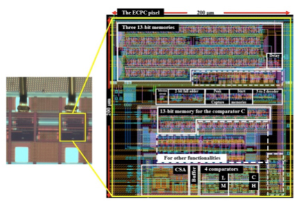
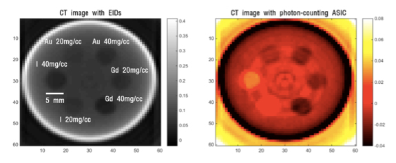
To develop a photon-counting spectral CT, one of the most important components is a photon-counting ASIC of which a fast signal processing ability to treat flux X-ray photons and a low noise performance are required. We have been developing a photon-counting ASIC with a new pulse-shape technique for a fast processing time. Both analog circuits and digital circuits necessary in processing X-ray photon signals are integrated in an area of 200 x 200 m2 as shown in Figure 1. The chip can be used up to X-ray fluence of 109 photons/mm2, which is the typical level used in a conventional CT. It consumes only 7.2 W/pixel and uses four energy bins to measure the energy information of incident X-ray photons into four colors or four materials. Recently a photon-counting spectral CT image for a test phantom was obtained using this chip and a CdTe detector shown in Figure 2. In the near future, when this colorful CT is available, a revolution in X-ray imaging can be achieved.
Cho, Gyuseong (Department of Nuclear and Quantum Engineering)
Homepage: http://radiation.kaist.ac.kr
E-mail: gscho@kaist.ac.kr
