


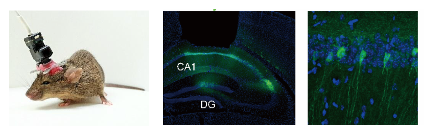
Prof. Jin-Hee Han’s group at KAIST has identified neural activity patterns associated with memory recall by recording the activity of a large number of neurons in the brain region called the hippocampus, essential for memory formation and recall, of freely moving mice during memory recall. A subpopulation of cells in the CA1 area of the hippocampus increased its firing responses by learning, and this change was significantly correlated with memory strength during recall. At the cell population level, synchronous cell activity patterns were associated with memory recall.
“Our findings are the first to show that the activity patterns of individual neurons and synchronously firing cell ensembles are involved in memory recall,” said Prof. Han, the corresponding author and an associate professor in the Department of Biological Sciences at the KAIST Institute for the Biocentury.
It has long been thought that memories are encoded and stored in sparse groups of neurons across broadly distributed areas in the brain. These cells are hypothetically referred to as engrams, where memory traces are thought to be allocated. Over the last decade, such engram cells have been experimentally demonstrated and precisely identified in animal studies. Despite these recent advances in identifying memory engrams, however, little is known about the activity patterns of individual neurons (i.e., electrical firing rate of brain cells) or cell ensembles that are correlated with memory recall.
“It has been technically very challenging to precisely record individual spikes and keep tracking them over days from low firing neurons as hippocampal CA1 neurons in freely behaving mice. It took us a lot of time and effort to establish the endoscope in vivo calcium imaging set-up in mice. To solve this problem, we decided to employ a recently developed calcium indicator, GCaMP7f, to provide better detection of individual spikes, and an advanced cell sorting algorithm, CNMF_E, for calcium image data. Luckily, it worked, leading us to successfully identify the neural activity patterns involved in memory recall,” said first author Dr. Han-Sol Lee, a graduate student in the Department of Biological Science at KAIST.
“Smaller subsets of neurons or even individual neurons can have distinct roles in encoding or retrieval of memory. Future advancements in techniques that enable precise manipulation of activity of individually targeted neurons in animals, combined with simultaneous recording of neuronal activity within networks, will address the causal role of hippocampal neurons with distinct activity patterns for encoding and retrieval of memories,” said Prof. Han.
From this study, the team observed that a subset of neurons located in the dorsal part of hippocampal CA1 increased their responses to learned context and that the amount of change was correlated with behavioral performance during memory recall. Importantly, these increases in response were specific to memory recall and not simply due to animal movement or immobility. At the ensemble level, synchronous cell activity patterns were associated with memory retrieval. Together, these results suggest that increases in responses of individual neurons and synchronous cell activity patterns in the dCA1 neuronal network are critically involved in representing contextual memory recall.
This work was supported by grants from Samsung Science and Technology Foundation (Project Number SSTF-BA1801-10).
Prof. Jin-Hee Han, Dr. Han-Sol Lee Dept. of Biological Sciences, KAIST
E-mail: Han.jinhee@kaist.ac.kr
Homepage: https://sites.google.com/site/neuralcircuitandbehaviorlab/
