The collaborative research team of Professor YongKeun Park and Professor Yong Jeong in KI for Health Science and Technology (KIHST) demonstrated depth-enhanced imaging of a living mouse tissue using wavefront shaping optical coherence tomography (WS-OCT).
Optical coherence tomography (OCT) is a noninvasive imaging tool for biological tissues. Based on low coherence interferometry, the reflected signals from samples are utilized for reconstructing tomographic images of tissues. However, the optical inhomogeneity of tissues disturbs the light propagation, or light distribution, inside tissues. This multiple light scattering prevents deep tissue imaging, and a typical penetration depth is roughly limited to 1 mm.
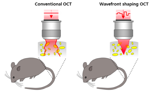
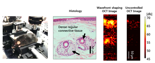
The research team has developed a new method called wavefront shaping optical coherence tomography or WS-OCT. This method adjusts the light beam by using a micro-mirror array and effectively controls the multiple scattering in the spectral domain of OCT system. Compared with the conventional OCT imaging, WS-OCT technique can enhance the penetration depth up to 92%. The researchers also demonstrated that WS-OCT increases the penetration depth in a mouse tail imaging in vivo.
Because the wavefront shaping enhances the penetration depth in various types of tissues, WS-OCT system could eventually lead to in vivo deep tissue imaging for diagnostic purposes. This work was published online in Journal of Biomedical Optics in February 2016.
Park, YongKeun (Associate Professor, Department of Physics)
Jeong, Yong (Associate Professor, Department of Bio and Brain Engineering)
Homepage: http://bmol.kaist.ac.kr, http://ibrain.kaist.ac.kr
E-mail: yk.park@kaist.ac.kr, yong@kaist.ac.kr