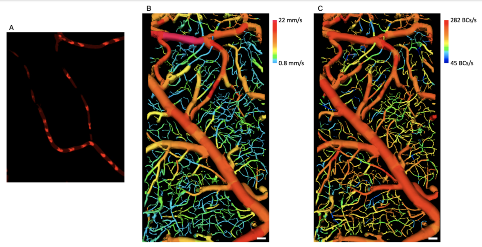
Prof. Wang-Yuhl Oh’s group in KI Health Science and Technology has developed, for the first time, an imaging technology that visualizes blood cells flowing in complex three-dimensional vascular networks without using any contrast agent.
This work was published on 11 October, 2023 in Small (Direct Blood Cell Flow Imaging in Microvascular Networks, Small 19(41), 2302244 (2023)).
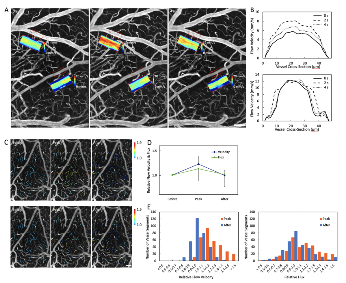
Various hemodynamic information about blood flow within microvascular networks is closely linked to the health of associated organs, making accurate measurement and analysis crucial for various disease studies. The ideal way to obtain the hemodynamic information would be directly imaging blood cells flowing within various vessels with high temporal resolution. However, to date, such technology does not exist and methods such as measuring other values correlated with blood flow velocity or injecting fluorescently labeled blood cells for imaging thus have been used instead. It is also challenging to selectively image only the blood cells due to the abundant reflected and scattered light from the surrounding tissues. A newly developed technique has garnered significant attention by directly imaging the flowing blood cells within the complex vascular networks across a wide three-dimensional area at high speed (acquiring 1,450 images per second) without using any exogenous substances such as fluorescent contrast agents.
Professor Oh’s research team has successfully visualized only the flowing blood cells by devising image processing methods based on the characteristics of flowing blood cells. Additionally, they prevented blood cells from being obscured by speckle noise by using spatially incoherent illumination. They also utilized a high-speed camera capable of acquiring large amounts of photons per pixel to enhance the imaging sensitivity, allowing flowing blood cell imaging from deep within the tissue.
Professor Oh remarked, “Various hemodynamic information such as blood flow velocity and blood cell flux within various vessels is crucial in biomedical research. While it would be best to directly image blood cells flowing at various speeds within blood vessels, such imaging devices or methods do not yet exist. Therefore, methods such as measuring Doppler signals related to blood flow velocity or imaging stained blood plasma or portions of blood cells using fluorescence microscopy have been primarily used. The newly developed technology enables rapid direct imaging of blood cells flowing within various vessels without injecting any contrast agent, making it not only very convenient for use but also enabling the immediate acquisition of accurate hemodynamic information.”
This work was supported by the National Research Foundation of Korea (2020R1A2C3009667) and the National Institutes of Health (3P41EB015903).


Prof. Wang-Yuhl Oh, Gyounghwan Kim Dept. of Mechanical Engineering, KAIST
E-mail: woh1@kaist.ac.kr
Homepage: https://phil.kaist.ac.kr


