Heart attacks, most frequently caused from the failure of the coronary artery, which supply blood to heart muscles, are the leading cause of death in industrialized countries. Recent developments in high-speed, second-generation (2G) Optical Coherence Tomography (OCT) enable the endoscopic microscopy of patients’ long coronary artery segments. This opens up a new horizon in the diagnostics of high-risk coronary plaques and hopefully leads to the development of new therapeutic methods.
Although OCT is the only imaging method that can provide sufficient resolution to visualize important microscopic features associated with high-risk coronary lesions, true high-resolution 3D visualization has not been achieved for long-coronary segments because longitudinal-spatial resolution is several times poorer than the cross-sectional resolution due to insufficient imaging speed.
Prof. Wang-Yuhl Oh’s research group recently developed an ultrahigh-speed intravascular OCT (IV-OCT) system and demonstrated the first true high-resolution 3D visualization of microstructures in the vessel walls in vivo. The development of a high-speed 2G-OCT system miniaturized optical catheters (endoscopic probes), and a high-speed and high-precision fiber-optic rotary junction enables high-speed IV-OCT imaging that is 3-8 times faster than the fastest current IV-OCT system.
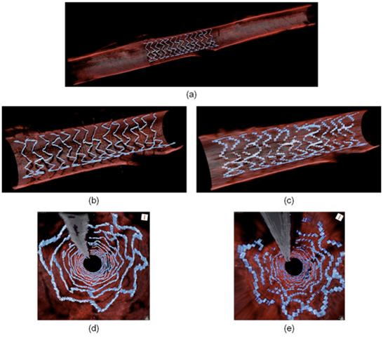
Images of rabbit aortae, obtained by the newly-developed high-speed IV-OCT system (Figure 1 (a), (b), and (d)), visualize microstructures in the vessels, such as the stent strut microstructure and the orifice of the side branch, much better than the ones imaged at the fastest previous speed (Figure 1 (c) and (e)). In the newly-developed system’s images with the conventional longitudinal pitch, it only takes one second to capture images of a 7.2-cm-long segment of a vessel.
Prof. Oh’s group is currently in the process of in-vivo coronary artery imaging for large animals, such as swine, and is planning to conduct clinical patients’ coronary artery imaging afterward.
This work was supported in part by the NRF of Korea, grant 2010-0017465, and by the MSIP of Korea, grant GFP/(CISS-2012M3A6A6054200), and was published on January 1, 2014 in Biomed Opt Express.