


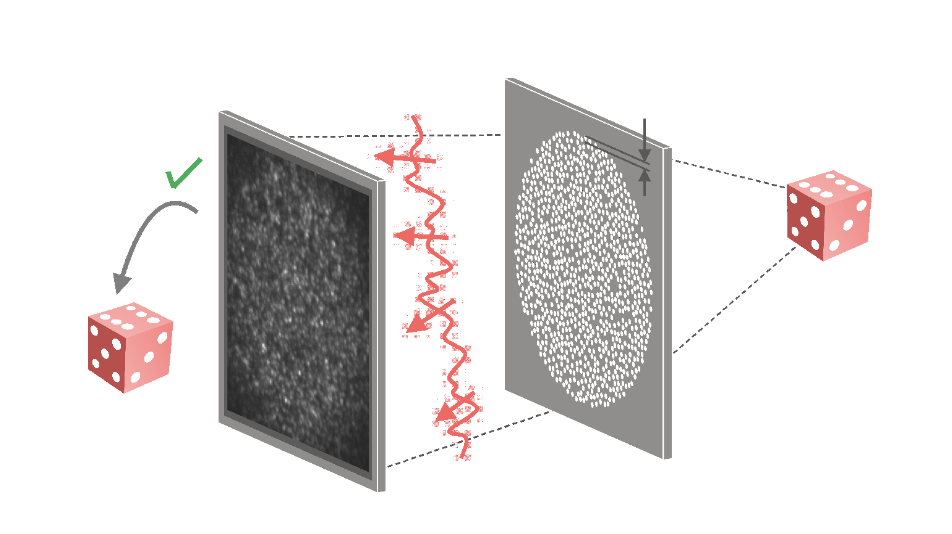
Prof. YongKeun Park’s group at KAIST has developed two advanced X-ray phase imaging methods in succession. They first achieved randomness-based X-ray imaging by introducing an X-ray diffuser after the samples. This technique allows them to directly retrieve a high-resolution phase image of the sample from a single speckle pattern, based on the mathematical rules of random vectors. They obtained a better spatial resolution (14 nm) than the feature size of the diffuser that was used (300 nm). This is a significant advantage over conventional X-ray imaging optics (zone plate) with a resolution limited by their feature size. This work was published on April 6, 2023 in [“Direct high-resolution X-ray imaging exploiting pseudorandomness.” Light: Science & Applications 12.1 (2023): 88]. In collaboration with Pohang Accelerator Laboratory (PAL), this new imaging system was applied to characterize the pulse-to-pulse field variations of self-amplified spontaneous emission (SASE) pulses from an X-ray free-electron laser (XFEL). The complex spatial features of the pulses were easily obtained from a single-pulse measurement and their statistics were analyzed. This work was published on March 8, 2023 in [“Pulse-to-pulse field characterization at x-ray free-electron lasers using a speckle-correlation scattering matrix.” Optica 10.3 (2023): 393-400].
“Our next step will be to pursue this approach. Our ultimate goal is to achieve sub-nanometer image resolution using X-rays,” said Dr. KyeoReh Lee, the first and corresponding author and a postdoctoral fellow in the Department of Physics at KAIST. “We believe that utilizing next-generation X-ray light sources, such as diffraction-limited storage rings, along with high-performance X-ray detectors, could enable us to surpass the current resolution record achieved through zone-plate-based imaging,” he added.
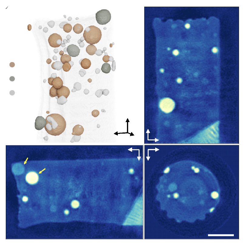
The second phase imaging method they proposed is even simpler. They managed to retrieve the quantitative phase by inserting a small piece of silicon wafer in the conventional X-ray microscopy. It partially blocks the sample diffraction and provides the phase image directly from a single image, using powerful mathematical equations called Kramers–Kronig (KK) relations. They successfully reconstructed the three-dimensional (3-D) refractive index distribution of mixed nanopowder, silicon, and integrated circuit samples. This work was published on March 9, 2023 in [“Full-field quantitative X-ray phase nanotomography via space-domain Kramers–Kronig relations.” Optica 10.3 (2023): 407-414].
“Using the developed nanotomography method, we can measure the 3-D electron density of nanomaterials with almost the same configuration as existing technology. This means that it can be easily reproduced at many accelerator X-ray sources around the world and used for 3-D structure and property analysis of various nanomaterials,” said Dr. KyeoReh Lee. “We expect that its performance will be enhanced when combined with the coherent X-rays that will be provided by the 4th generation synchrotron radiation accelerator that will be newly established in Ochang, Chungbuk,” he added.
This work was supported by Tomocube, KAIST Advanced Institute for Science-X, National Research Foundation of Korea (2015R1A3A2066550, 2021R1C1C2009220, 2022M3H4A1A02074314), an Institute of Information & Communications Technology Planning & Evaluation (IITP) grant funded by the Korean government (MSIT) (2021-0-00745), and the Technology Innovation Program (20011661) funded by the Ministry of Trade, Industry & Energy (MOTIE).
Prof. YongKeun Park, Dr. KyeoReh Lee Department of Physics, KAIST
E-mail: yk.park@kaist.ac.kr
Homepage: http://bmol.kaist.ac.kr
