Functional hyperemia in the rat cortex was investigated using high-speed optical coherence tomography (OCT) angiography. OCT angiography (OCTA) was performed to image the hemodynamic stimulus-response over a wide field of view. Temporal changes in vessel diameters in different vessel compartments were measured in order to monitor localized hemodynamic changes. This research demonstrates the potential of OCTA for the investigation of neurovascular coupling in small animal models.

The research team of Prof. Wang-Yuhl Oh in KI for Health Science and Technology (KIHST) investigated the sensory-evoked hemodynamic changes in the rat barrel cortex using optical coherence tomography (OCT) to demonstrate the potential of high-speed OCT for the investigation of neurovascular coupling in the rodent brain. Sensory-evoked changes in both vessel diameter and capillary hemodynamics were monitored over a wide field of view using OCT angiography (OCTA), as shown in Figure 1. The results showed prominent dilation of downstream arterioles with modest dilation of upstream arteries, which are several hundreds of micrometers away from the arterioles. On the other hand, veins show relatively negligible dilation.
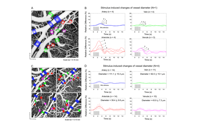
Figure 1. Investigation of the rapid hemodynamic response using OCTA. (A) An en face OCT angiogram with the regions where vessel diameters are measured indicated with colored boxes. Blue, red, green, and magenta color boxes represent arterial, arteriolar, venous, and venular segments, respectively. (B) Changes in vessel diameters as a function of time for the vessels indicated in A. (C) En face OCT angiograms of four different animals. (D) The average relative hemodynamic responses for the four vessel compartments.

Temporal changes of the OCTA signal in the capillary bed were also investigated using the same data. An increase in the signal in capillaries can be interpreted as an overall increase in capillary blood flow.

Figure 2. Investigation of the hemodynamic changes in capillaries. (A) Regions of the capillary bed were manually segmented on the en face angiogram. The regions close to the activation site are shaded in red and the regions on the periphery are shaded in blue. (B) The mean changes in the signals for the regions close to the activation site. (C) Spatiotemporal changes in the signals in capillaries (in Animal 2). Capillaries that show more than a 3% increase in the signals are shaded in blue.

In summary, both spatial and temporal changes in diameters of various vessel compartments were simultaneously monitored using OCTA. The results of this study suggest that OCT can be highly useful in the investigation of neurovascular coupling in the rodent brain.

This work was recently accepted for publication in Journal of Cerebral Blood Flow and Metabolism (JCBFM).

Contact Information:
Prof. Wang-Yuhl Oh (Department of Mechanical Engineering)
Paul Shin (Department of Mechanical Engineering)
Homepage: http://bpil.kaist.ac.kr
E-mail: woh1@kaist.ac.kr