


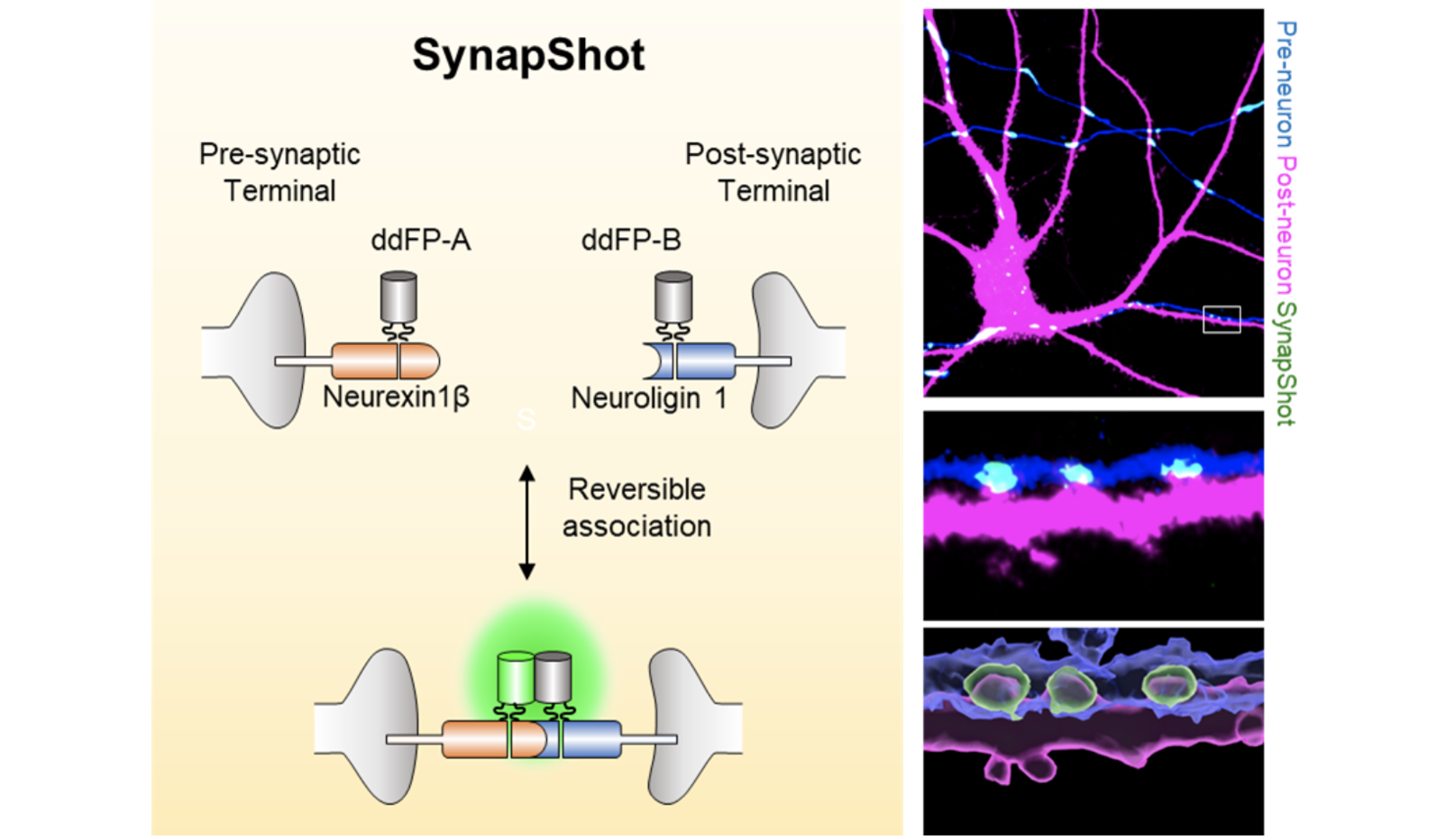
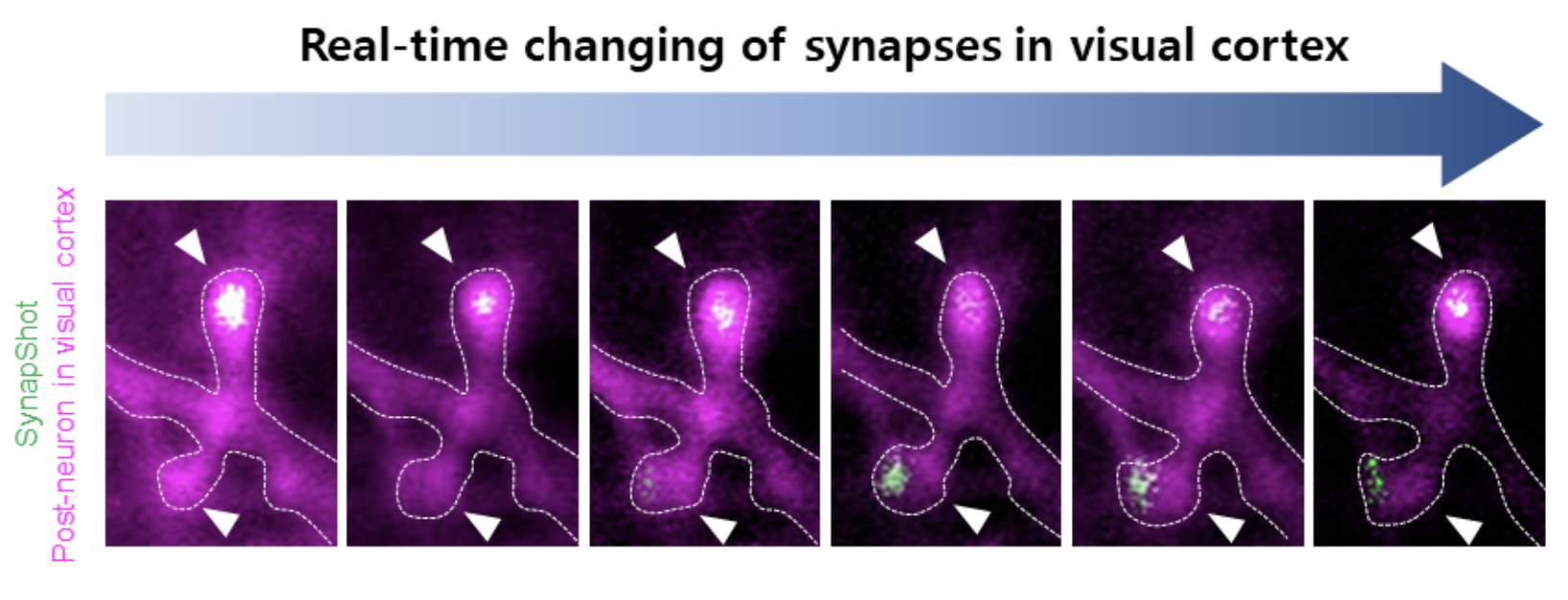
Prof. Won Do Heo’s group at KAIST has developed ‘SynapShot,’ a method for visualizing the structural dynamics of intact synapses by combining dimerization-dependent FPs (ddFPs) with engineered synaptic adhesion molecules. SynapShot allows real-time monitoring of reversible and bidirectional changes of synaptic contacts under physiological stimulation. The application of green and red ddFPs in SynapShot enables simultaneous visualization of two distinct populations of synapses. Notably, the red-shifted SynapShot is highly compatible with blue light-based optogenetic techniques, allowing for visualization of synaptic dynamics while precisely controlling specific signaling pathways. Furthermore, we demonstrate that SynapShot enables real-time monitoring of structural changes in synaptic contacts in the mouse brain during both primitive and higher-order behaviors. The study was published on 8 January 2024 (Real-time visualization of structural dynamics of synapses in live cells in vivo, Nature Methods, 21, 353–360 (2024).
SynapShot is more specialized for real-time visualization of synaptic dynamics in live cells and the intact brain, complementing split GFP-based methodologies. Overall, this platform will contribute to our understanding of the role of time-dependent changes in the synaptic structure under diverse physiological and pathological conditions in the brain.
“Through our collaborative research with international research teams, synapse shot technology has opened up the possibility of directly observing the rapid and dynamic formation and changes of synapses, which was difficult to realize in the past. This technology is expected to revolutionize the research methodology in the field of neuroscience research and play an important role in the future of brain science,” said Won Do Heo, the corresponding author and a professor in the Department of Biological Sciences and the KAIST Institute for the Biocentury at KAIST.
“This technique is significant because it is the first in the world to visualize the formation and removal of synapses in real time and because it can label intact synapses more accurately than existing techniques,” said the first author Seungkyu Son, a graduate student.
This work is based on research that has been conducted as part of the KAIST-funded Global Singularity Research Program for 2022 (W.D.H.). It was also supported by a National Research Foundation of Korea grant funded by the Korean government (MSIT) (no. 2020R1A2C301474213, W.D.H.)
Prof. Won Do Heo, Dr. Seungkyu Son, Dr. Jinsu Lee Dept. of Biological Sciences, KAIST
E-mail: wondo@kaist.ac.kr
Homepage: https://sites.google.com/view/heolab
