


Prof. Wang-Yuhl Oh’s group in KI Health Science and Technology has developed, for the first time, a cellular-level high-resolution imaging technology in a wide field human retina, not only at the focal plane but also at all three-dimensional locations outside the focal plane.
This work was published on March 15, 2023 in Small (Wide-Field Three-Dimensional Depth-Invariant Cellular-Resolution Imaging of the Human Retina, Small 19(11), 2203357 (2023)).
Cell-level resolution imaging of the retina is essential for early diagnosis of diseases and enhancing our understanding of retinal diseases. Because the retina must be imaged through the lens of the eye, high-resolution imaging is challenging due to aberrations (e.g., astigmatism) of the eye lens. Previously, to overcome this problem, researchers have developed an adaptive optics method that uses optical hardware to measure the aberration of the eye lens and correct it. However, this method not only requires complex and expensive additional optical hardware, but also allows high-resolution imaging only at a single focal plane. In order to obtain three-dimensional high-resolution image data, imaging should be performed repeatedly at multiple depths by changing the focal position.
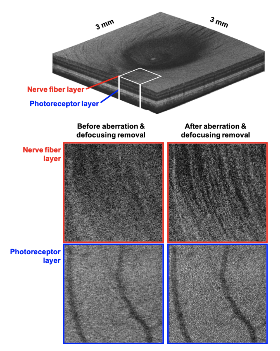
The newly developed technology acquires 3D retinal image data at once using a simple standard imaging optics and computationally removes aberrations and defocusing, which causes image blur outside the focal plane. Prof. Oh said, “For computational aberration and defocusing removal, the intensity of light scattered from each 3D position on the retina as well as the phase value must be accurately known.” He added, “Because the amount of data (the number of pixels constituting the image) for cellular-resolution imaging of a wide 3D retina becomes very large, an imaging technology that can acquire 3D image data at ultra-high speed is essential, and this means an ultra-fast phase-stable 3-D imaging system is essential.” The newly developed OCT imaging system has solved the phase instability issue of existing OCT technologies, while providing imaging speed that is more than 20 times faster than the current fastest commercial retinal OCT system, enabling 3D imaging of the human retina spanning 3 mm x 3 mm (consisting of approximately 10 billion 3D pixels) at the cellular level in 2.3 seconds.
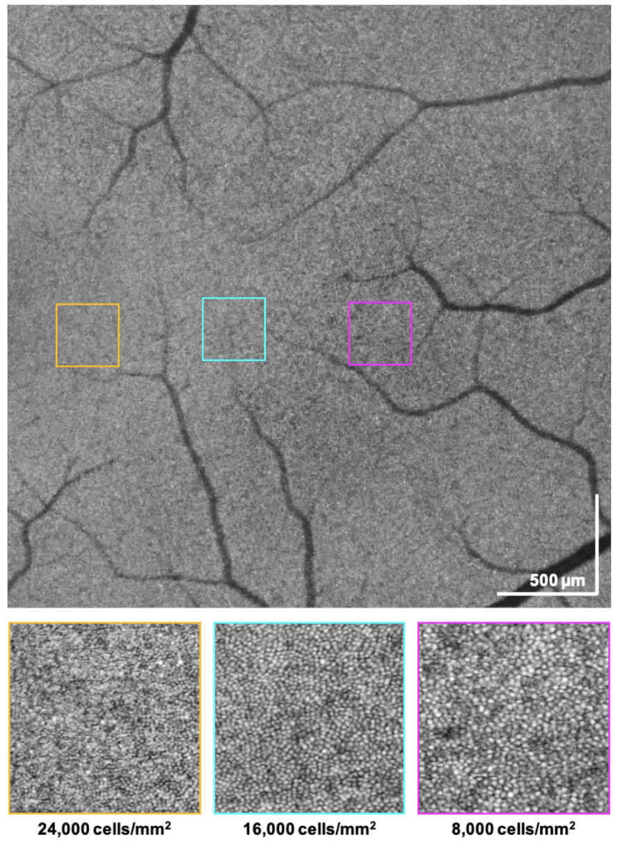
Prof. Oh also emphasized, “While using the same standard imaging optics as the retinal OCT systems currently used in hospitals, the new technology provides visualization of the wide-field 3D cellular-level structures in various retinal layers, such as the retinal nerve fibers and photoreceptor cells. This should be very useful in both clinical and research fields of retinal diseases.
This work was supported by the National Research Foundation of Korea (2020R1A2C3009667) and the National Institutes of Health (3P41EB015903).
Prof. Wang-Yuhl Oh, Dr. ByunKun Lee Dept. of Mechanical Engineering, KAIST
E-mail: woh1@kaist.ac.kr
Homepage: https://phil.kaist.ac.kr
