-
Research Highlight
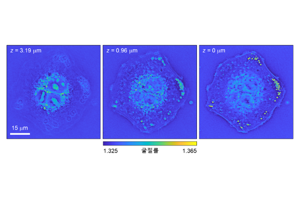
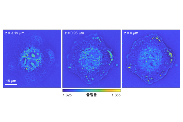
3D microanatomy of unlabeled cancer tissues revealed by holotomography and virtual H&E
Prof. Park’s group at KAIST developed 3D virtual H&E staining of thick cancer tissues using holotomography and AI. ...read more
-
Research Highlight
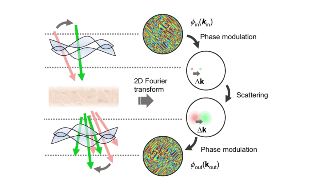
Digital aberration correction for enhanced thick tissue imaging exploiting a aberration matrix and tilt-tilt correlation from the optical memory effect
KAIST researchers develop high-resolution label-free imaging technology for thick biological tissues...read more
-
Research Highlight
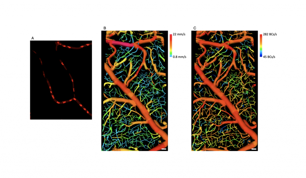
Direct Blood Cell Flow Imaging in Microvascular Networks
Prof. Wang-Yuhl Oh’s group has developed, for the first time, an imaging technology that visualizes blood cells flowing in complex three-dimensional vascular networks without using any contrast agent. ...read more
-
Research Highlight Top Story
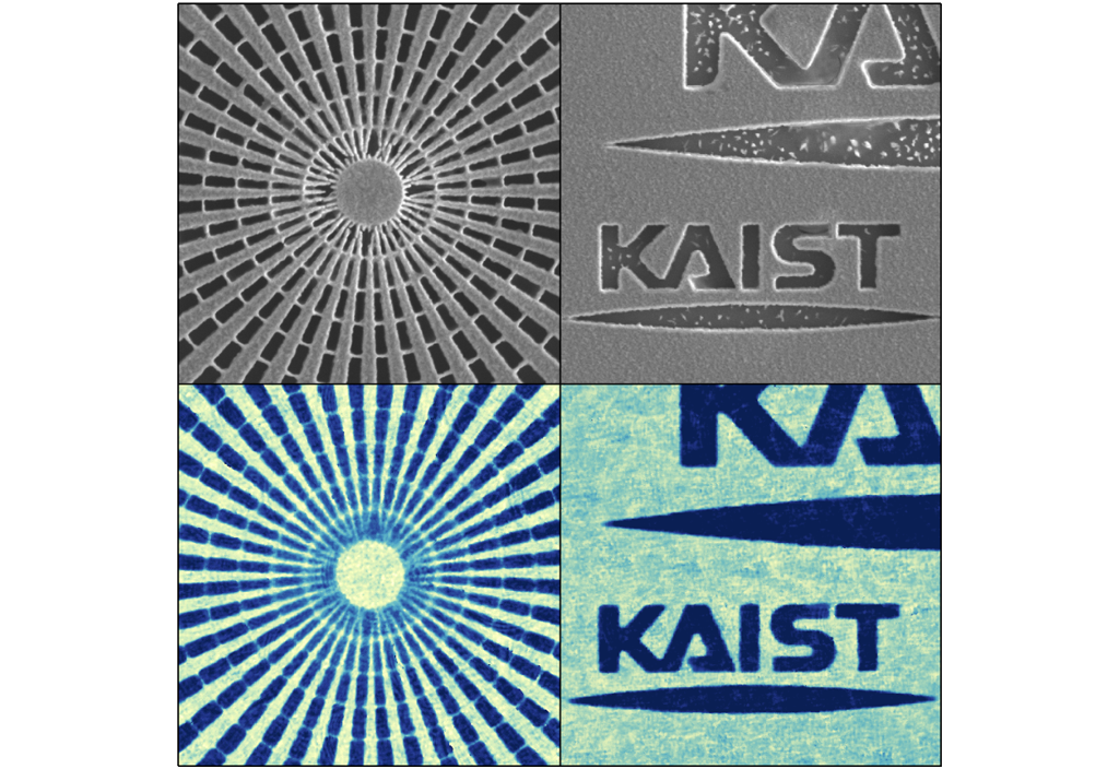
High-resolution quantitative X-ray phase nanoimaging based on cutting-edge optical imaging methods
Prof. Park’s group has developed a novel high-resolution quantitative X-ray phase imaging system that can overcome two long-standing challenges in X-ray nanoimaging: the limitations in image resolution and the instability of phase retrieval methods. They applied optical imaging techniques that they have recently developed in the same group. The imaging systems have been successfully tested in both synchrotron source and X-ray free-electron laser facilities at Pohang Accelerator Laboratory....read more
-
Research Highlight
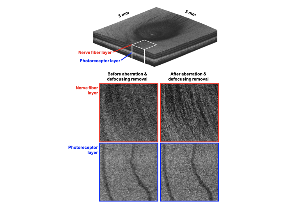
Wide-Field Three-Dimensional Depth-Invariant Cellular-Resolution Imaging of the Human Retina
Prof. Wang-Yuhl Oh’s group has developed, for the first time, a cellular-resolution imaging technology in a wide field human retina at all three-dimensional locations....read more
-
Research Highlight
New holographic microscopy enables the 3D label-free imaging of biological specimens without interferometry
Prof. YongKeun Park’s group has developed a novel microscopy technique for the label-free imaging of biological specimens. Based on Kramers-Kronig relations, this new method produces quantitative phase images of biological specimens with an optical microscope simply by controlling the incident angle of illumination....read more
-
Research Highlight
New method enables holographic imaging without interferometry
Prof. YongKeun Park’s group has developed a novel method for non-interferometric holographic imaging. The method, based on Kramers-Kronig relations, provides holographic images of biological specimens using an optical microscope simply by controlling the illumination beam....read more
-
Research Highlight
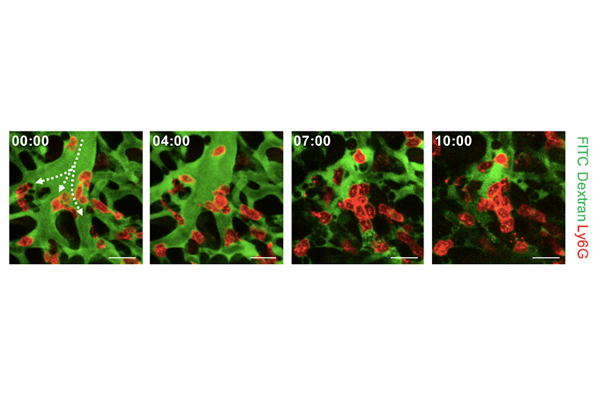
Uncovering the Dead Space of Pulmonary Microcirculation in Sepsis
An innovative bio-imaging technique enables real-time in vivo visualization of pulmonary microcirculation, leading to the discovery of active cluster formation of neutrophils inside the microvasculature during the pathogenesis of acute lung injury in sepsis...read more
-
Research Highlight
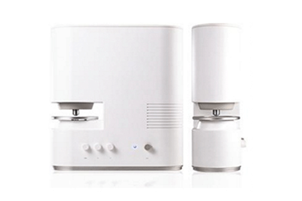
KAIST Start-up Set to Release Tomographic Microscope for 3D Live Cell Imaging
3-D Holographic microscopy from a KAIST start-up was launched and enables scientists to visualize cells and tissues without labeling...read more

291 Daehak-ro Yuseong-gu Daejeon, 34141, Republic of Korea
Partnered with KAIST Breakthroughs and KAIST Compass

