-
Research Highlight
Quantitative hemodynamic analysis of cerebral blood flow and neurovascular coupling using optical coherence tomography angiography
Functional hyperemia in the rat cortex was investigated using high-speed optical coherence tomography (OCT) angiography. OCT angiography (OCTA) was performed to image the hemodynamic stimulus-response over a wide field of view. Temporal changes in vessel diameters in different vessel compartments were measured in order to monitor localized hemodynamic changes. This research demonstrates the potential of OCTA for the investigation of neurovascular coupling in small animal models....read more
-
Research Highlight
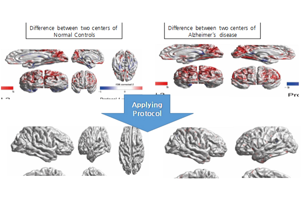
A novel method for improving T1 MR image compatibility
A novel method can improve T1 MR image compatibility across different centers to study Alzheimer's disease ...read more
-
Research Highlight
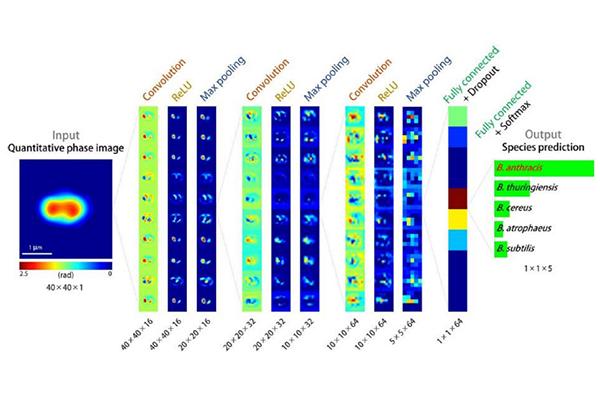
Rapid Detection of Anthrax Spores using Artificial Intelligence and Holographic Microscopy
Combining holography with deep learning enables rapid optical screening of anthrax spores as well as other pathogens....read more
-
Research Highlight
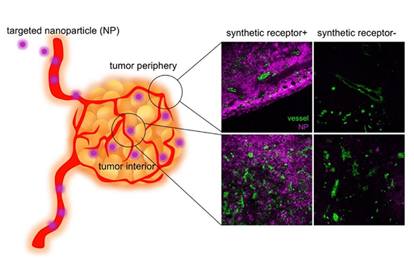
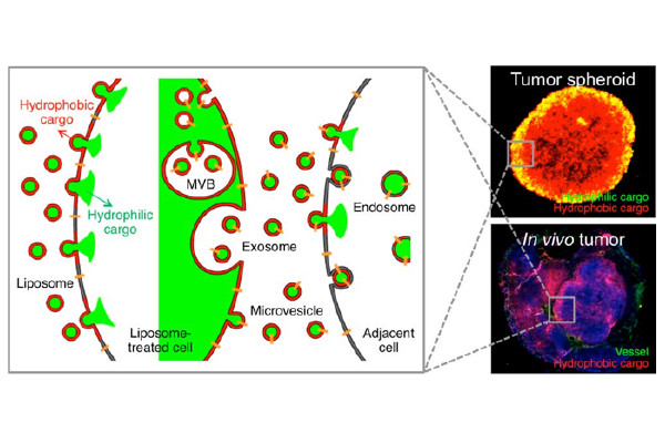
Decorating Tumor Cell Membranes with Synthetic Receptors to Improve Targeted Cancer Therapy
Delivery of synthetic receptors to tumor cell membranes using both synthetic and biological nanoparticles led to effective treatment of cancer....read more
-
Research Highlight
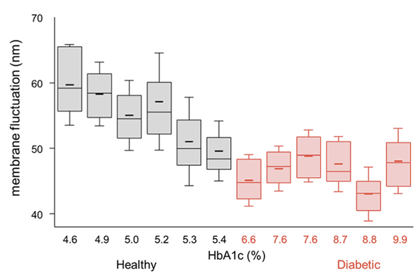
Observing Changes in Diabetic Red Blood Cell Properties Using Non-invasive 3-D Holographic Microscopy
Alterations in morphological, biochemical, and mechanical properties of individual diabetic red blood cells were quantitatively and non-invasively studied employing a 3-D quantitative phase imaging technique. No significant alterations were observed in the morphological and biochemical parameters of diabetic red blood cells compared to the healthy ones. However, membrane fluctuations of the diabetic red blood cells were significantly diminished and exhibited negative correlation with HbA1c levels, implying a deteriorated deformability of diabetic RBCs....read more
-
Research Highlight
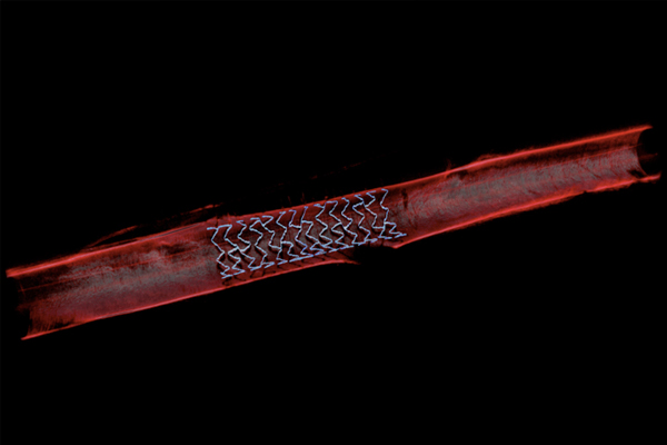
A Depth-Enhanced Method in Deep Tissue Imaging of a Living Mouse
A new method called wavefront shaping optical coherence tomography (WS-OCT) enables in vivo deep tissue imaging of a mouse....read more

291 Daehak-ro Yuseong-gu Daejeon, 34141, Republic of Korea
Partnered with KAIST Breakthroughs and KAIST Compass